X-rays are an essential part of modern dentistry, offering clear and precise imaging of the teeth, jawbones, and surrounding structures. They are used for both diagnostic and treatment planning purposes in children and adults. Whether evaluating the eruption of teeth in young patients or detecting deep decay or bone loss in adults, dental X-rays provide critical insights that cannot be seen during a visual examination. These images guide everything from routine check-ups to complex surgical planning. With advancements in digital technology, dental radiography is now faster, safer, and more accurate.
Pediatric patients benefit from low-dose radiation systems specifically designed for smaller anatomy, while adults benefit from detailed imaging that supports preventive and restorative care. From routine bitewings to advanced 3D scans, X-rays play a crucial role in delivering high-quality, customized dental care. They ensure early detection, accurate diagnosis, and effective treatment outcomes across all age groups.
RVG Imaging
RVG (Radiovisiography) is an advanced digital intraoral imaging technology that has revolutionized dental diagnostics with its speed, accuracy, and reduced radiation exposure. This system provides high-resolution images in real-time, allowing dental professionals to immediately view and evaluate the internal structure of teeth and surrounding bone. Used extensively for routine checkups, root canal evaluations, cavity detection, and post-treatment monitoring, RVG is a reliable diagnostic tool for both children and adults.
- Enhanced Safety and Speed for Kids and Adults: RVG reduces radiation by up to 80%, offering a safer diagnostic option, especially for children, without compromising image clarity or speed of delivery.
- Detailed Imaging for Accurate Diagnosis: This technology allows dentists to see minute changes in bone and tooth structure, aiding in early detection of issues such as decay, infection, or root abnormalities in adults and children.
- Ergonomic and Patient-Friendly Design: The small, rounded digital sensor fits comfortably in all mouth sizes, providing a smoother experience for children and adults with sensitive gag reflexes.
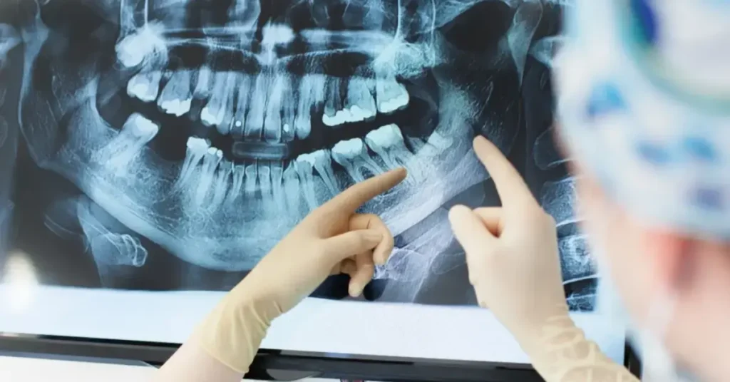
OPG X-Ray
An Orthopantomogram (OPG) is a panoramic scanning dental X-ray that displays a complete image of the mouth, including all teeth, both jaws, the temporomandibular joints (TMJ), and surrounding structures in a single frame. It plays a vital role in diagnosing and evaluating dental and skeletal conditions that may not be fully visible through intraoral X-rays. Suitable for both children and adults, an OPG offers a non-invasive and efficient method for screening multiple areas of the oral cavity at once.
- Wide-Field Image for Comprehensive Evaluation: This single scan offers a broad view of the entire oral structure, helping identify cysts, fractures, or bone loss affecting several teeth at once.
- Ideal for Monitoring Growth and Development: In children and adolescents, OPG helps monitor tooth eruption patterns, spacing, and jawbone development, making orthodontic planning more precise.
- Quick, Comfortable, and Non-Invasive: Since there’s no need to place a sensor inside the mouth, the process is comfortable even for young children or adults with dental anxiety or limited mouth opening.
CBCT Scan
Cone Beam Computed Tomography (CBCT) is a highly advanced imaging technique that generates three-dimensional views of dental, skeletal, and soft tissue structures with unmatched precision. Unlike traditional two-dimensional X-rays, CBCT provides a complete cross-sectional analysis of the jaw, teeth, nerves, and sinuses, making it an essential tool for comprehensive diagnosis and treatment planning in both children and adults. This technology is especially valuable in complex cases where exact positioning and measurement are critical, such as implant placement, impacted teeth evaluation, or jaw joint assessment.
- Three-Dimensional Accuracy for Complex Cases: CBCT provides a precise view of bone density, nerve location, and sinus anatomy, critical for planning implants or assessing abnormalities.
- Pediatric Assessment with Greater Clarity: For children, it helps identify impacted teeth, jaw asymmetry, and developmental concerns without the need for multiple traditional X-rays.
- Efficient and Low-Dose Imaging: The scan takes less than a minute and targets only the required area, using focused radiation to keep the procedure safe and fast for all age groups.
Digital Dental X-Rays
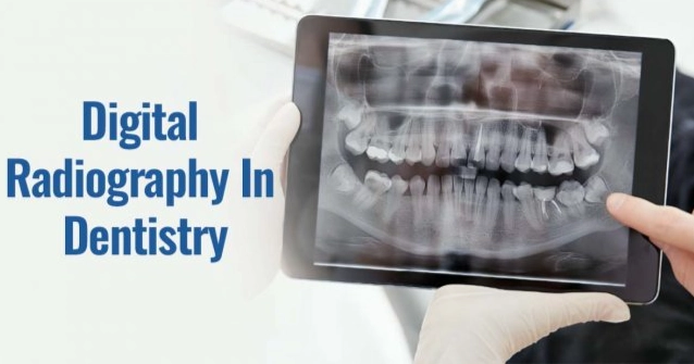
Digital dental X-rays are a modern imaging solution that has replaced traditional film-based radiographs in many dental practices. This technology provides crisp, clear images almost instantly, greatly enhancing diagnostic accuracy and reducing wait times for both children and adults. Whether it’s a routine check-up or part of a more detailed treatment plan, digital X-rays offer reliable insights into the condition of the teeth, bone, and surrounding tissues. Because the system uses sensors instead of film, the process is faster, more comfortable, and involves far less radiation exposure, which is especially beneficial for pediatric patients and those requiring frequent imaging.
- Real-Time Imaging for Faster Decisions: Digital X-rays are displayed instantly, enabling dentists to explain findings to patients during the same visit and begin treatment without delay.
- Minimal Radiation Exposure: Children and adults benefit from significantly lower radiation, allowing for more frequent monitoring without health concerns over cumulative exposure.
- Environmentally Friendly and Easily Shared: Digital files reduce chemical waste and can be sent to specialists electronically, making them ideal for interdisciplinary care involving orthodontics, periodontics, or oral surgery.
Are dental x-rays are safe and needed?
Yes, dental X-rays are generally safe and are often necessary for diagnosing and monitoring oral health conditions that are not visible during a regular dental exam.
Safety of Dental X-rays
Low Radiation Exposure:
- Modern dental X-ray technology uses very low levels of radiation. The amount of radiation from a typical dental X-ray is much lower than that from other medical imaging techniques.
- For example, a single dental X-ray exposure has a very small dose of radiation, roughly equivalent to the radiation exposure you would receive from natural background sources over a few days.
Precautionary Measures:
- Lead Apron & Thyroid Collar: Dentists use protective gear like lead aprons and thyroid collars to shield vital organs and reduce radiation exposure to sensitive areas.
- Digital X-rays: Many dental offices now use digital X-ray systems, which emit even less radiation compared to traditional film-based X-rays and provide faster, more accurate results.
Why Dental X-rays Are Needed
- Detect Hidden Problems: X-rays allow the dentist to detect problems that may not be visible during a regular checkup. This includes cavities between teeth, tooth infections, bone loss, and issues with the roots of teeth.
- Monitor Oral Health Progress: X-rays help monitor the progress of dental treatments, such as the placement of fillings, crowns, or braces. They also track the health of teeth after procedures like root canals or extractions.
- Diagnose Jawbone and Gum Disease Issues: They are essential for detecting conditions like periodontal disease (gum disease) or bone loss caused by infections or other oral health problems, which can impact the health of your teeth and gums.
- Assist in Planning Treatments: Dentists often use X-rays when planning for more complex procedures like implants, braces, or tooth extractions, ensuring they understand the full structure of your teeth, jaw, and bones.
- Early Detection of Oral Cancer: X-rays can sometimes help identify signs of oral cancer or tumors in the early stages, which may not be immediately apparent during a visual exam.
Dental X-rays are safe, necessary, and crucial for detecting hidden dental issues, planning treatments, and maintaining overall oral health. The risk of radiation exposure is minimal, especially with modern techniques and protective measures, and the benefits of early detection and treatment typically far outweigh any potential risks. Your dentist will ensure that X-rays are only taken when necessary for your specific needs.
Full Mouth X-Ray
A full mouth X-ray series (FMX) is a detailed collection of intraoral radiographs that provide a comprehensive view of every tooth, its roots, and the surrounding bone structures. Typically consisting of multiple periapical and bitewing images, FMX is a foundational diagnostic tool in dentistry. It is particularly valuable during a patient’s first visit or when planning for extensive dental treatments, including orthodontics, implants, and full-mouth rehabilitation. Both children and adults benefit from this imaging technique, as it offers a complete picture of oral health in a single session.
- Complete View for Thorough Diagnosis: It reveals hidden decay, root infections, bone abnormalities, or signs of gum disease that may not be visible during a routine dental exam.
- Monitors Development in Growing Children: Helps evaluate tooth eruption patterns, spacing issues, and alignment in children, allowing early intervention in orthodontic or growth-related concerns.
- Essential for Adults with Complex Needs: Provides a full understanding of the oral condition for patients requiring implants, dentures, or treatment for advanced periodontal disease, ensuring nothing is overlooked.
Conclusion
X-rays are a key part of modern dental care, offering accurate and detailed images for both children and adults. From RVG and OPG to CBCT and full-mouth scans, each method helps detect hidden issues and supports precise treatment planning. Children benefit from fast, low-radiation scans, while adults receive comprehensive evaluations for long-term care and restorative needs.At Dr.Hari’s Dental Clinic, we use advanced imaging systems combined with expert analysis to ensure accurate diagnosis and personalized care. Our technology-driven approach improves comfort, safety, and treatment outcomes. Whether it’s a child needing orthodontic evaluation or an adult preparing for surgery, our imaging supports timely, effective care. With the right diagnostic tools, we help patients of all ages maintain healthier, more confident smiles.