Enamel and dentin defects are a range of dental issues that significantly impact the strength, appearance, and functionality of teeth. These defects can occur in both children and adults due to various factors such as genetic disorders, developmental problems, nutritional deficiencies, systemic illnesses, or environmental factors. In children, enamel and dentin defects can affect primary teeth or developing permanent teeth, leading to problems like sensitivity, decay, and difficulties with chewing and speaking. In adults, these defects may have long-term effects, leading to worn-down teeth, cracks, or increased vulnerability to cavities and infections. Early detection and treatment of enamel and dentin defects are critical to preventing complications such as tooth loss, misalignment, or discomfort.
Management of Enamel Defects
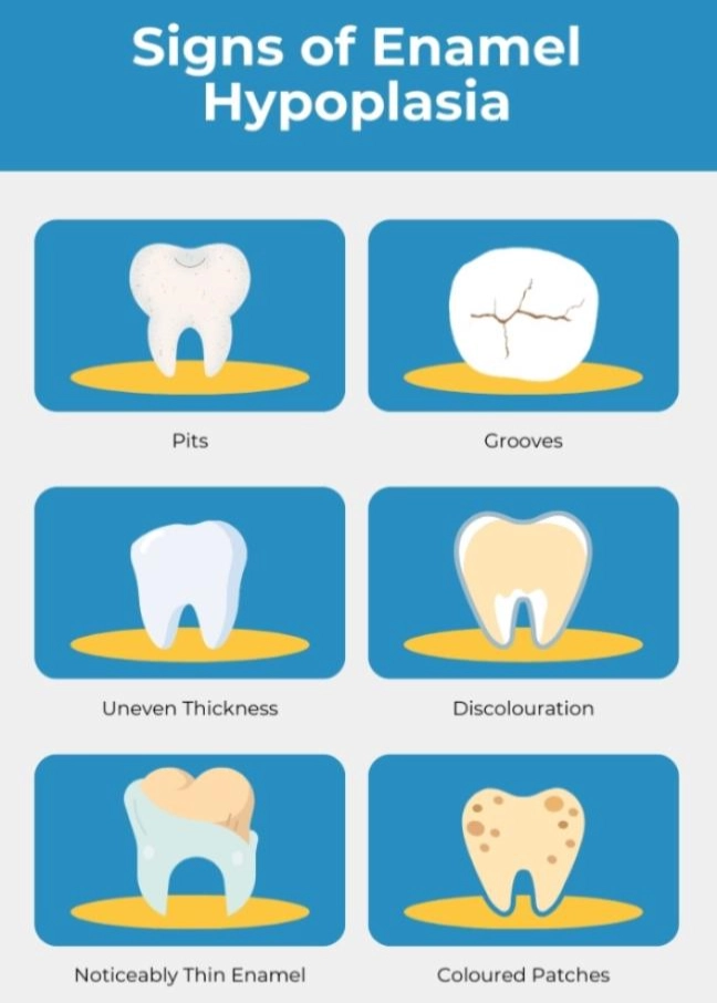
Enamel Hypoplasia
Enamel hypoplasia is characterized by underdeveloped or thin enamel, resulting in weak, discolored, or pitted teeth. It may arise due to genetics, systemic illness, or trauma during tooth development. Common in both primary and permanent dentition, it increases susceptibility to tooth decay, particularly in children.
- Early Diagnosis and Restoration: Timely diagnosis allows for customized treatment strategies. Composite fillings or crowns help restore function, reduce sensitivity, and protect the remaining tooth structure.
- Preventive Care: Fluoride application, a low-sugar diet, and good oral hygiene can strengthen enamel-deficient areas and lower decay risk in children and adults.
- Functional and Aesthetic Solutions: In adults, options such as porcelain veneers, bonded restorations, or ceramic crowns improve appearance and reinforce enamel, ensuring both aesthetics and function.
Enamel Hypomineralisation
Enamel hypomineralisation is a defect where enamel is poorly mineralized, leading to soft, porous, and weak tooth structure. This condition results in teeth with white, yellow, or brown spots, and can affect the chewing surface, especially the first molars and incisors. In children, this condition is often associated with developmental issues and can make teeth more prone to decay and wear. Early detection and intervention are essential to prevent further damage and ensure effective treatment.
- Minimally Invasive Intervention: For mild cases, resin infiltration and sealants reduce sensitivity and halt damage while preserving tooth structure.
- Monitoring and Protective Coverage: Teeth with significant mineral loss may need full-coverage restorations like stainless steel crowns for children and ceramic crowns for adults to restore strength and function.
- Ongoing Evaluation: Routine dental visits and radiographs help monitor enamel stability, allowing treatment adjustments to maintain comfort and function.
Dental Fluorosis
Dental fluorosis is caused by excessive fluoride consumption during tooth development, leading to discoloration and surface irregularities on enamel. The severity of fluorosis ranges from mild, with white streaks or spots, to severe, where the enamel is pitted or discolored with brown stains. In children, it’s important to monitor fluoride use during critical periods of tooth formation, such as when they are younger than six. In adults, fluorosis is mostly a cosmetic concern, but severe cases may require cosmetic restorative procedures like microabrasion, bleaching, or veneers.
- Cosmetic Treatment Options: Microabrasion and bleaching improve aesthetics in mild to moderate cases. Severe cases may require porcelain veneers or full ceramic crowns to restore a uniform appearance.
- Preventive Education: Educating parents on correct fluoride usage pea-sized toothpaste, avoiding swallowing, and monitoring fluoride sources helps reduce the risk of fluorosis in children.
- Comprehensive Management: Severe fluorosis may require cosmetic bonding, high-quality veneers, or crowns to restore function and aesthetics.
Amelogenesis Imperfecta
Amelogenesis imperfecta (AI) is a genetic disorder that affects the enamel, resulting in thin, soft, or absent enamel, leading to increased sensitivity, wear, and susceptibility to decay. This condition can affect both primary and permanent teeth and may vary in severity depending on the type of AI. In children, the condition can affect both baby and adult teeth, causing issues with chewing and speech. Depending on the severity of the defect, treatment may include the use of crowns, composite restorations, or veneers to restore the affected teeth.
- Customized Treatment Plans: Depending on the AI type, treatment may include crowns, composite restorations, or veneers to improve structure and appearance
. - Multidisciplinary Car: Children benefit from a combined approach including restorative, orthodontic, and preventive treatments to manage function and aesthetics long-term.
- Supportive Care and Counseling: Parents need guidance on managing dental sensitivity, nutrition, and psychosocial aspects such as self-esteem and speech development.
Molar Incisor Hypomineralisation (MIH)
Molar Incisor Hypomineralisation (MIH) affects the first permanent molars and incisors, causing white spots, brown stains, or defective enamel on the affected teeth. It is a developmental defect that can cause significant sensitivity and increased susceptibility to cavities, especially on molars. Children with MIH may experience pain, difficulty chewing, and cosmetic concerns. In adults, MIH may cause significant functional and aesthetic problems in their molar and incisor teeth, requiring restorative treatments.
- Early Recognition and Pain Management: Prompt diagnosis enables the use of desensitizing agents and minimally invasive restorations, ideal for children.
- Preventive Resin Sealants: High-viscosity resin sealants protect weak enamel from bacterial invasion and acids, delaying further decay.
- Restorative and Orthodontic Planning: Severe cases may require full-coverage restorations. Orthodontic assessments help manage eruption delays or alignment issues caused by compromised molars.
Dentin Defects Management
Dentin Dysplasia
Dentin dysplasia is a genetic disorder that affects the formation of dentin, leading to weakened tooth structure and an increased risk of decay and fracture. This condition can cause the teeth to appear normal externally but have short roots and fragile structures internally. In children, the primary concern is ensuring that the teeth remain functional as long as possible, with regular monitoring and preventive care. In adults, restorative procedures like crowns or root canal therapy may be required if the teeth become infected or damaged. A multidisciplinary approach, including orthodontics and prosthodontics, is often needed to manage long-term complications.
- Radiographic Diagnosis and Monitoring: Radiographs reveal internal structural anomalies such as short roots and pulp calcification. Ongoing monitoring is essential to detect complications early.
- Functional Rehabilitation: Treatment includes root canal therapy followed by crowns or prosthetic restorations to restore strength and function while preserving the tooth.
- Preventive Emphasis: Fluoride varnishes, professional cleanings, and excellent oral hygiene are crucial in preserving affected teeth and minimizing secondary infections.
Regional Odontodysplasia
Regional odontodysplasia is a developmental anomaly characterized by the abnormal formation of enamel, dentin, and pulp in a localized area. This condition often presents as a hypoplastic tooth with an enlarged pulp chamber and poorly developed roots. It typically affects one or a few teeth and is most commonly found in the maxillary region. Treatment depends on the severity of the defect and may involve extractions, restorative procedures, or orthodontic care to maintain function and aesthetics.
- Clinical and Radiographic Features: Teeth appear yellowish or brown and radiographically resemble “ghost teeth” due to large pulp chambers and thin dentin and enamel layers.
- Individualized Treatment Approach: Treatment varies by severity. Temporary restorations or space maintainers may be used in children. In adults, prosthetic replacements such as crowns or implants are considered after growth is complete.
- Multistage Care Planning: Due to frequent eruption delays or absence, long-term care involving pediatric dentists, prosthodontists, and orthodontists is necessary for functional and aesthetic restoration.
Conclusion
Effective diagnosis and management of enamel and dentin defects are crucial for preserving oral health in both children and adults. From conditions like dental fluorosis to complex disorders like amelogenesis imperfecta and regional odontodysplasia, individualized care ensures better outcomes, improved function, and enhanced appearance. At Dr. Hari’s Dental Centre, we use advanced diagnostics and personalized care to manage these dental challenges. Our expert team is committed to restoring comfort, confidence, and long-term oral health helping every patient achieve a strong, healthy smile.
Read also: General Dentistry


